abumaizar
administrator | 19 December, 2023
Navigating severely calcified canals in dentistry can be a real headache. It’s like dealing with a tricky puzzle where traditional methods often fall short.
This article is your guide through the maze of tough cases, shedding light on the problems we face with the usual approaches. We’re here to shake things up, waving goodbye to the old ways, and shouting from the rooftops about the promising solution that guided endodontics brings to the table.
As we unfold this story, you’ll see how guided endodontics swoops in to save the day.
Severely calcified canals present unique challenges for clinicians. These canals are obstructed by mineralized deposits, making it difficult to locate, clean, and shape the root canal system effectively.
Guided endodontics offers a promising solution for clinicians faced with the intricacies of calcified canal management.
Incorporating advanced imaging techniques, such as cone beam computed tomography (CBCT), and navigational instruments, guided endodontics enables precise treatment planning and real-time visualization, enhancing accuracy and reducing treatment time.
Understanding Calcified Canals and Endodontic Complexity
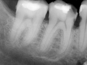
Calcified canals refer to root canals that have become hardened and obstructed due to the deposition of calcified tissue.
These calcifications can occur naturally as a result of aging or secondary to trauma.
Endodontic complexity arises when calcified canals present challenges to accessing and navigating through the root canal system. The calcifications can vary in density and location, making it difficult for clinicians to effectively clean and shape the canals and complete the root canal treatment.
Additionally, calcified canals can be curved, narrow, or have irregular anatomy, further complicating treatment.
Accessing and navigating calcified canals require advanced skills and specialized instruments. Standard endodontic files may not be effective in removing the calcifications, which can hinder the successful completion of root canal therapy.
Introduction to Guided Endodontics
Guided endodontic procedures have evolved over time, thanks to advancements that have made them more accessible and effective.
The Rise of Guided Endodontic Procedures
Guided endodontics has gained significant recognition in recent years as a revolutionary approach to root canal therapy. This innovative technique utilizes advanced imaging technology and computer-assisted planning to enhance treatment outcomes.
With guided endodontic procedures, clinicians can precisely navigate complex root canal anatomies and overcome the challenges presented by calcified canals. The integration of three-dimensional imaging, virtual treatment planning, and real-time navigation has revolutionized the way endodontic treatment is performed, offering clinicians enhanced accuracy, improved visualization, and reduced treatment times.
Advantages Over Traditional Endodontic Methods
The advantages of guided endodontics over traditional methods are promising. By adopting guided endodontic procedures, clinicians can overcome the limitations of conventional techniques and achieve more predictable treatment outcomes.
– Improved Accuracy: The use of 3D imaging technology allows clinicians to accurately plan and execute endodontic procedures, minimizing the risk of errors and improving the precision of treatment.
– Enhanced Visualization: Guided endodontics provides clinicians with a detailed and comprehensive view of the root canal system, enabling them to visualize and navigate intricate canal anatomy more effectively.
– Reduced Treatment Time: The integration of guided techniques streamlines the treatment process, allowing clinicians to work more efficiently and reducing the overall time required to complete the procedure.
– Predictable Outcomes: By combining advanced imaging, computer-assisted planning, and guided navigation, clinicians can achieve more predictable and successful treatment outcomes for patients.
The Role of CBCT

In guided endodontic treatment, cone beam computed tomography (CBCT) plays a crucial role in enhancing the precision and accuracy of the procedure.
CBCT imaging provides clinicians with detailed three-dimensional information about the root canal system, enabling them to develop effective treatment plans and navigate complex anatomical structures with confidence.
Unlike traditional two-dimensional imaging techniques (e.g. periapical radiographs), CBCT allows for a comprehensive evaluation of the teeth and surrounding structures in high resolution.
This detailed visualization enhances the clinician’s understanding of the canal anatomy, allowing them to identify calcifications, hidden canals, and other complexities that may affect treatment outcomes.
By incorporating CBCT into guided endodontics, clinicians can accurately assess the dimensions, position, and angulation of the canals, facilitating precise treatment planning. This imaging modality helps in determining the optimal access point, designing custom 3D printed guides, and selecting appropriate instruments for the procedure.
During treatment, CBCT imaging enables real-time navigation and verification, allowing the clinician to monitor the progress and make any necessary adjustments. This dynamic feedback ensures the accurate placement of files and irrigation solutions, reducing the risk of errors and complications.
Furthermore, CBCT imaging aids in post-treatment evaluation, enabling clinicians to assess the quality of the canal filling and ensure the absence of any untreated or missed canals.
This evaluation contributes to the long-term success of the endodontic treatment by identifying potential areas of concern and allowing for timely intervention.
Case Selection Criteria for Guided Endodontics
When managing severely calcified canals, it is important to carefully select cases that are suitable for guided endodontics.
Clinicians should consider factors such as the degree of canal calcification, the presence of any complex anatomical variations, and the overall prognosis of the tooth.
Custom 3D Printed Guides

Planning and precision are crucial factors that contribute to successful treatment outcomes. One innovative tool that enhances precision is the use of custom 3D printed guides. These guides are created based on accurate imaging data and treatment plans, ensuring a tailored approach for each patient.
Custom 3D printed guides provide clinicians with precise guidance during the endodontic procedure. They are designed to fit the patient’s unique anatomy, enabling accurate placement of instruments and ensuring the preservation of healthy tooth structure.
Using these custom 3D printed guides, clinicians can navigate the complexities of calcified canals with greater ease and precision, resulting in improved treatment outcomes.
Importance of Accurate Imaging
Accurate imaging plays a vital role in guided endodontics, enabling clinicians to visualize the intricate structures of calcified canals in detail. Cone beam computed tomography (CBCT) imaging provides clinicians with a three-dimensional view of the root canal system, allowing for precise treatment planning.
Incorporating accurate imaging techniques, clinicians can identify the extent of canal calcification and plan the most appropriate approach to navigate these challenging canal systems. Accurate imaging also aids in the identification of additional canals, anomalies, and potential complications that may arise during the procedure.
Combining Advanced Imaging with Navigational Instruments
The integration of advanced imaging techniques, such as cone beam computed tomography (CBCT), and intraoral scanners has transformed guided endodontics. CBCT imaging provides detailed three-dimensional information about the root canal system, enabling precise treatment planning and navigation.
This advanced imaging, combined with navigational instruments, allows clinicians to visualize the canal system in real-time and navigate it with unparalleled accuracy.
The use of intraoral scanners further enhances the integration of advanced imaging with navigational instruments. These scanners capture detailed digital impressions of the patient’s teeth and surrounding structures, which can be used to create virtual models for treatment planning.
Software Developments Enhancing Endodontic Treatment
In addition to advanced imaging and navigational instruments, software developments have played a crucial role in enhancing endodontic treatment. Virtual treatment planning software allows clinicians to simulate and visualize the entire endodontic procedure before it is performed on the patient.
This not only improves the accuracy of treatment planning but also helps clinicians anticipate potential challenges and develop effective strategies to overcome them.
Computer-aided design (CAD) software has also revolutionized the fabrication of custom surgical guides for guided endodontic procedures.
By digitally designing and manufacturing these guides, clinicians can ensure accurate transfer of their treatment plans to the clinical setting, further enhancing precision and reducing variability.
Simulation tools have also been developed to aid in the training and education of clinicians in guided endodontics. These tools allow aspiring clinicians to practice and refine their skills in a virtual environment, improving their confidence and competence before performing procedures on actual patients.
The integration of advanced imaging, navigational instruments, and software developments has transformed guided endodontic procedures, offering clinicians unprecedented accuracy and efficiency in the treatment of severely calcified canals. These technological advancements continue to push the boundaries of endodontic treatment, ensuring better outcomes and improved patient satisfaction.
Conclusion
In conclusion, guided endodontics plays a crucial role in managing severely calcified canals. Throughout this article, we have explored the comprehensive benefits of guided endodontics over traditional techniques. By combining precise treatment planning, advanced imaging, and navigational instruments, clinicians can effectively navigate the complexities associated with calcified canals and achieve predictable treatment outcomes.
With further advancements in technology and ongoing research, guided endodontics will continue to evolve and enhance the success of endodontic treatment for severely calcified canals. The integration of cone beam computed tomography (CBCT) and navigational instruments has revolutionized the field, allowing for accurate treatment planning and real-time visualization.


